Label the structures of the cochlea. – Embark on a scientific voyage to label the structures of the cochlea, the intricate organ responsible for our sense of hearing. This comprehensive guide delves into the anatomy, function, and significance of this remarkable structure, promising an auditory odyssey that will captivate your curiosity.
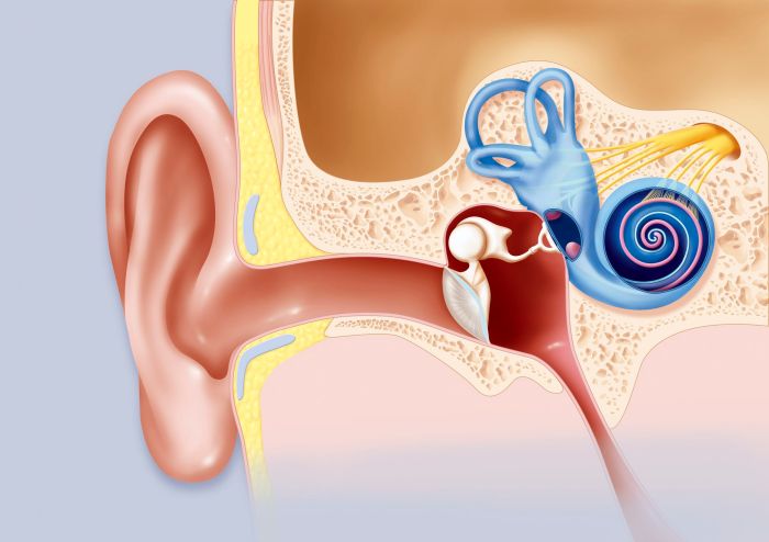
The cochlea, a marvel of nature’s design, resides within the inner ear, a testament to the wonders of human physiology. Its intricate architecture, a symphony of interconnected components, plays a pivotal role in our ability to perceive sound, transforming vibrations into electrical signals that our brains interpret as a symphony of melodies and harmonies.
Structures of the Cochlea: Label The Structures Of The Cochlea.

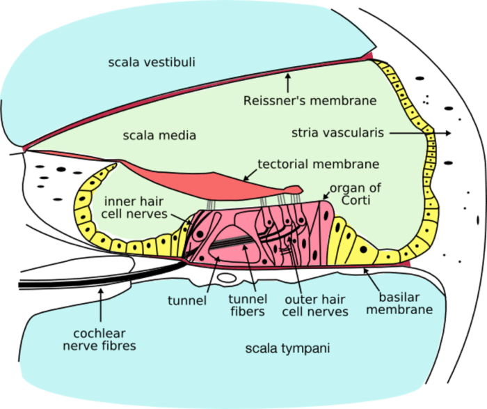
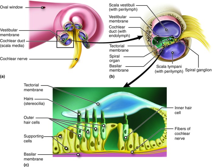
The cochlea is a spiral-shaped organ located in the inner ear that is responsible for hearing. It is divided into three chambers, or scalae, which are filled with fluid. The scala vestibuli is located above the scala media, and the scala tympani is located below the scala media.
The scala media is separated from the scala vestibuli by Reissner’s membrane, and from the scala tympani by the basilar membrane.
The basilar membrane is a thin, flexible membrane that vibrates in response to sound waves. The vibrations of the basilar membrane are then transmitted to the hair cells of the organ of Corti, which convert the vibrations into electrical signals that are sent to the brain.
Scala of the Cochlea
- Scala vestibuli:The scala vestibuli is located above the scala media and is filled with perilymph.
- Scala tympani:The scala tympani is located below the scala media and is also filled with perilymph.
- Scala media:The scala media is located between the scala vestibuli and the scala tympani and is filled with endolymph.
Reissner’s Membrane and Basilar Membrane
Reissner’s membraneis a thin, flexible membrane that separates the scala vestibuli from the scala media. It is made up of collagen and elastin fibers.
The basilar membraneis a thin, flexible membrane that separates the scala media from the scala tympani. It is made up of collagen and elastin fibers, and it is thicker and stiffer at the base of the cochlea than it is at the apex.
Organ of Corti
The organ of Corti is a complex structure located on the basilar membrane. It contains hair cells, which are the sensory cells of hearing. The hair cells are arranged in four rows, with the inner hair cells located closest to the scala media and the outer hair cells located closest to the scala tympani.
The hair cells are connected to the tectorial membrane, which is a gelatinous membrane that overlies the organ of Corti. When sound waves cause the basilar membrane to vibrate, the hair cells move in response to the movement of the tectorial membrane.
This movement causes the hair cells to release neurotransmitters, which are then sent to the brain.
Spiral Ganglion, Label the structures of the cochlea.
The spiral ganglion is a cluster of nerve cells located in the modiolus of the cochlea. The spiral ganglion cells are the first neurons in the auditory pathway. They receive signals from the hair cells of the organ of Corti and send them to the brain.
Top FAQs
What is the primary function of the cochlea?
The cochlea’s primary function is to transform sound vibrations into electrical signals, enabling us to perceive sound.
How many scalae are found within the cochlea?
There are three scalae within the cochlea: scala vestibuli, scala tympani, and scala media.
What is the role of the organ of Corti?
The organ of Corti, located within the cochlea, is responsible for converting sound vibrations into electrical signals, which are then transmitted to the brain.


